Plus d’un million de livres à portée de main !
Bookbot

Torsten B. Möller

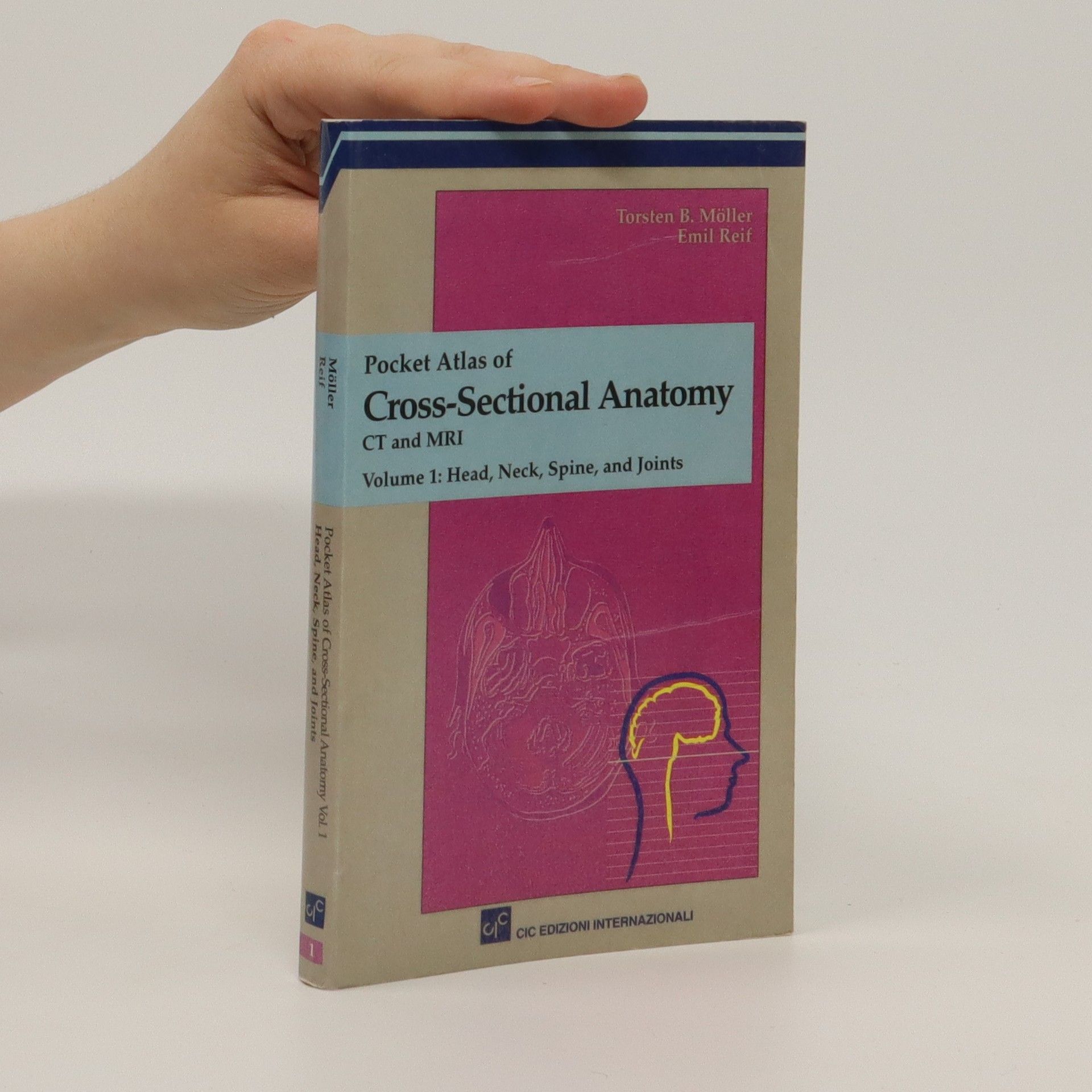
    Pocket atlas of cross sectional anatomy 1
    Anatomie IRM des membres
    Atlas des techniques radiologiques
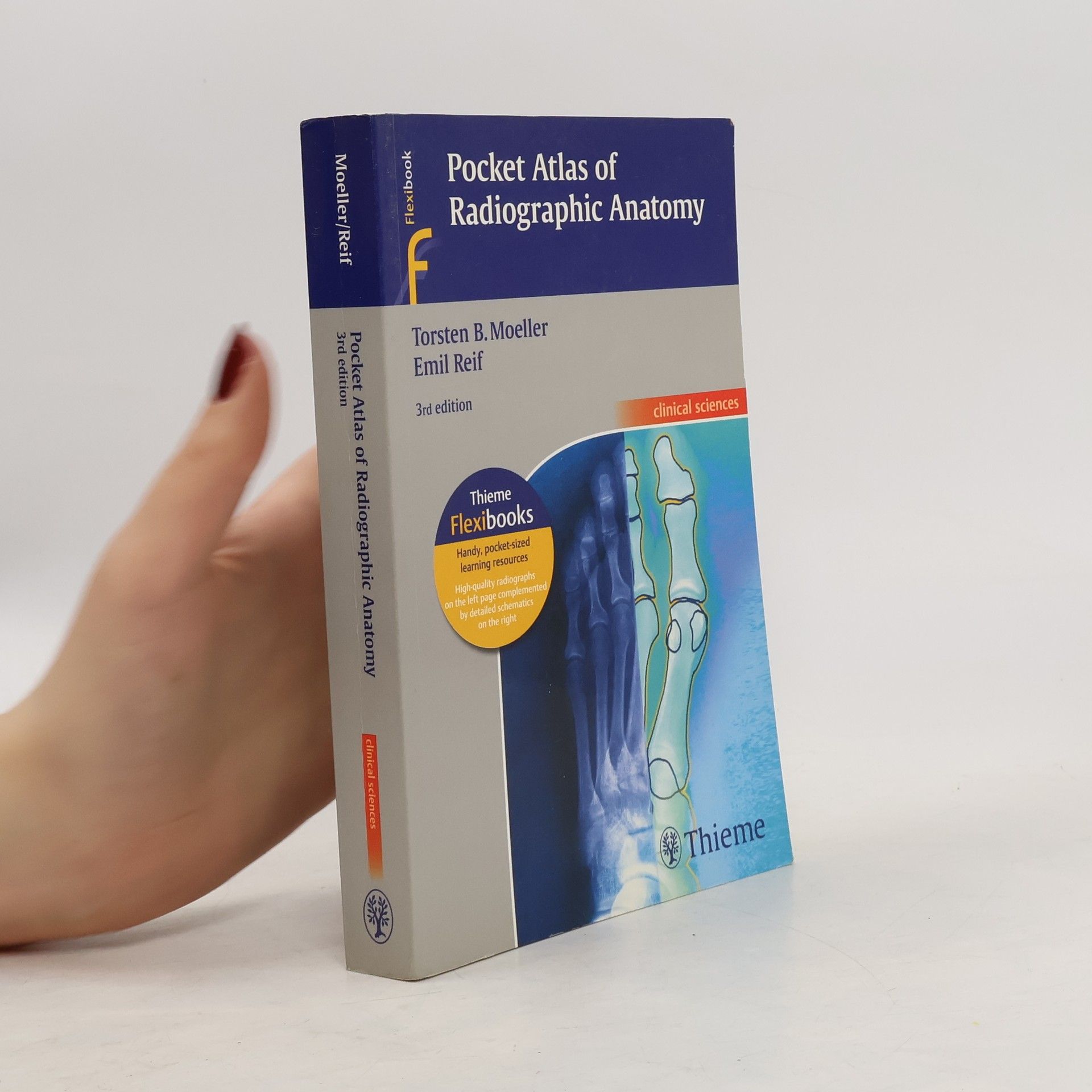
    Atlas de poche d'anatomie radiologique
    Atlas de poche d'anatomie en coupes sériées 1
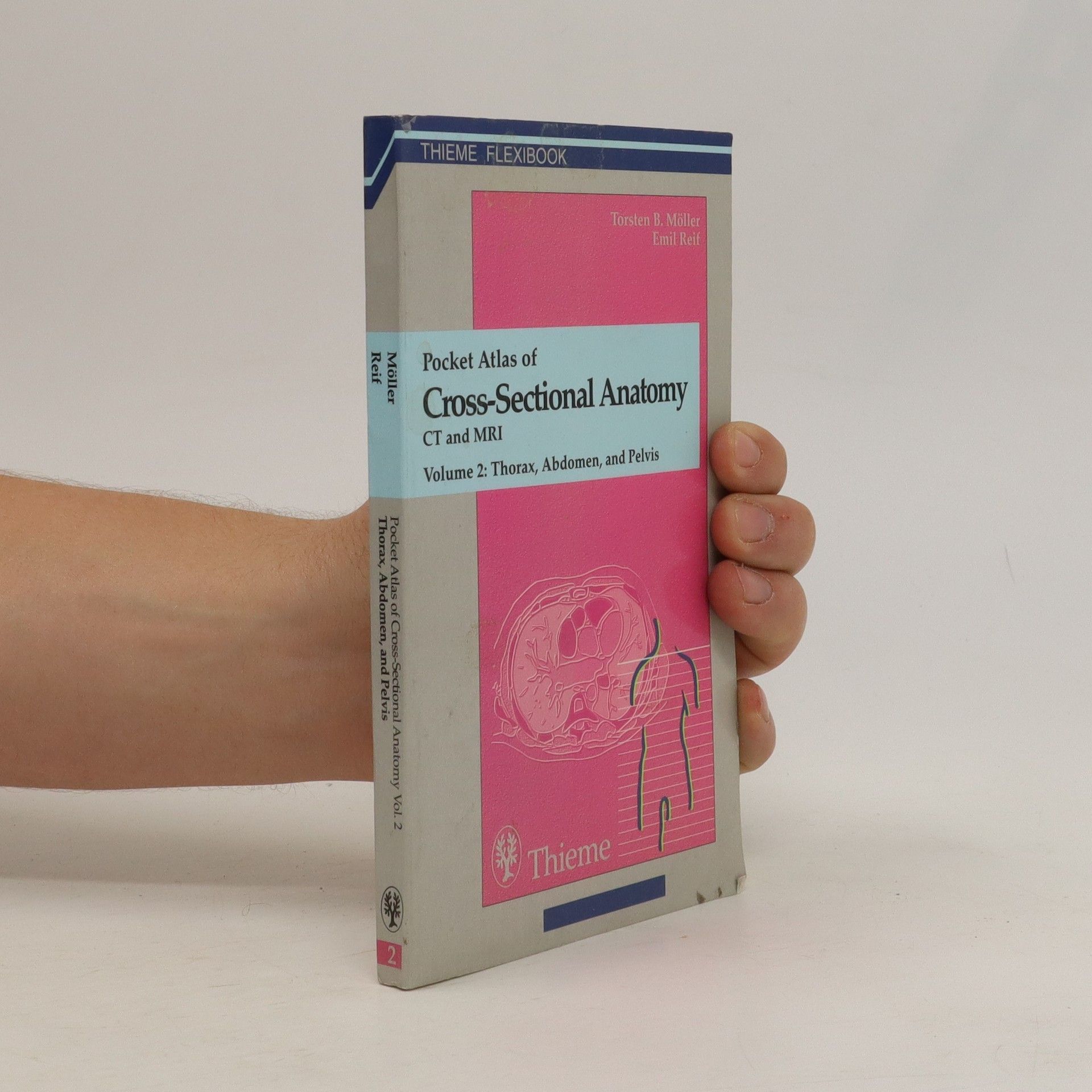
    Atlas de poche d'anatomie en coupes sériées 2
    • Cet Atlas de poche en deux volumes réussit un véritable pari pédagogique : il présente en effet, annoté de manière précise, actuelle et exhaustive, l'ensemble des structures anatomiques, jusque dans leurs plus infimes détails, visibles sur les images de tomodensitométrie et de résonance magnétique. Il constitue ainsi une référence et une aide précieuse à l'interprétation de ces images. Chacune des 250 planches radiologiques, intéressant les grandes régions (crâne, cou, rachis, thorax, abdomen, pelvis et articulations) est accompagnée d'un schéma sur lequel les différents systèmes anatomiques sont représentés par des couleurs-codes facilitant leur identification. Cet ouvrage s'adresse aux étudiants en radiologie, aux radiologues, à tous les médecins qui prescrivent des examens radiologiques ; il intéressera également tous les étudiants en médecine soucieux de parfaire leurs connaissances en anatomie humaine.

      Atlas de poche d'anatomie en coupes sériées 2
    • Cet Atlas de poche en deux volumes réussit un véritable pari pédagogique : Il présente en effet, annoté de manière précise, actuelle et exhaustive, l'ensemble des structures anatomiques, jusque dans leurs plus infimes détails, visibles sur les images de tomodensitométrie et de résonance magnétique. Il constitue ainsi une référence et une aide précieuse à l'interprétation de ces images. Chacune des 250 planches radiologiques, intéressant les grandes régions (crâne, cou, rachis, thorax, abdomen, pelvis et articulations) est accompagnée d'un schéma sur lequel les différents systèmes anatomiques sont représentés par des couleurs-codes facilitant leur identification. Cet ouvrage s'adresse aux étudiants en radiologie, aux radiologues, à tous les médecins qui prescrivent des examens radiologiques ; il intéressera également tous les étudiants en médecine soucieux de parfaire leurs connaissances en anatomie humaine.

      Atlas de poche d'anatomie en coupes sériées 1
    • Cet atlas de poche expose de façon claire et didactique l'anatomie radiologique normale nécessaire à l'interprétation des clichés radiographiques. Pour 170 examens radiographiques, un cliché standard, pris sous différentes incidences, est accompagné de sa reproduction dessinée au trait et légendée pour faciliter l'identification des structures anatomiques. L'utilisation de la terminologie radiologique et de la nomenclature anatomique nouvelle, ainsi que le rappel de la nomenclature traditionnelle dans l'index, font de cet ouvrage un outil essentiel.

      Atlas de poche d'anatomie radiologique
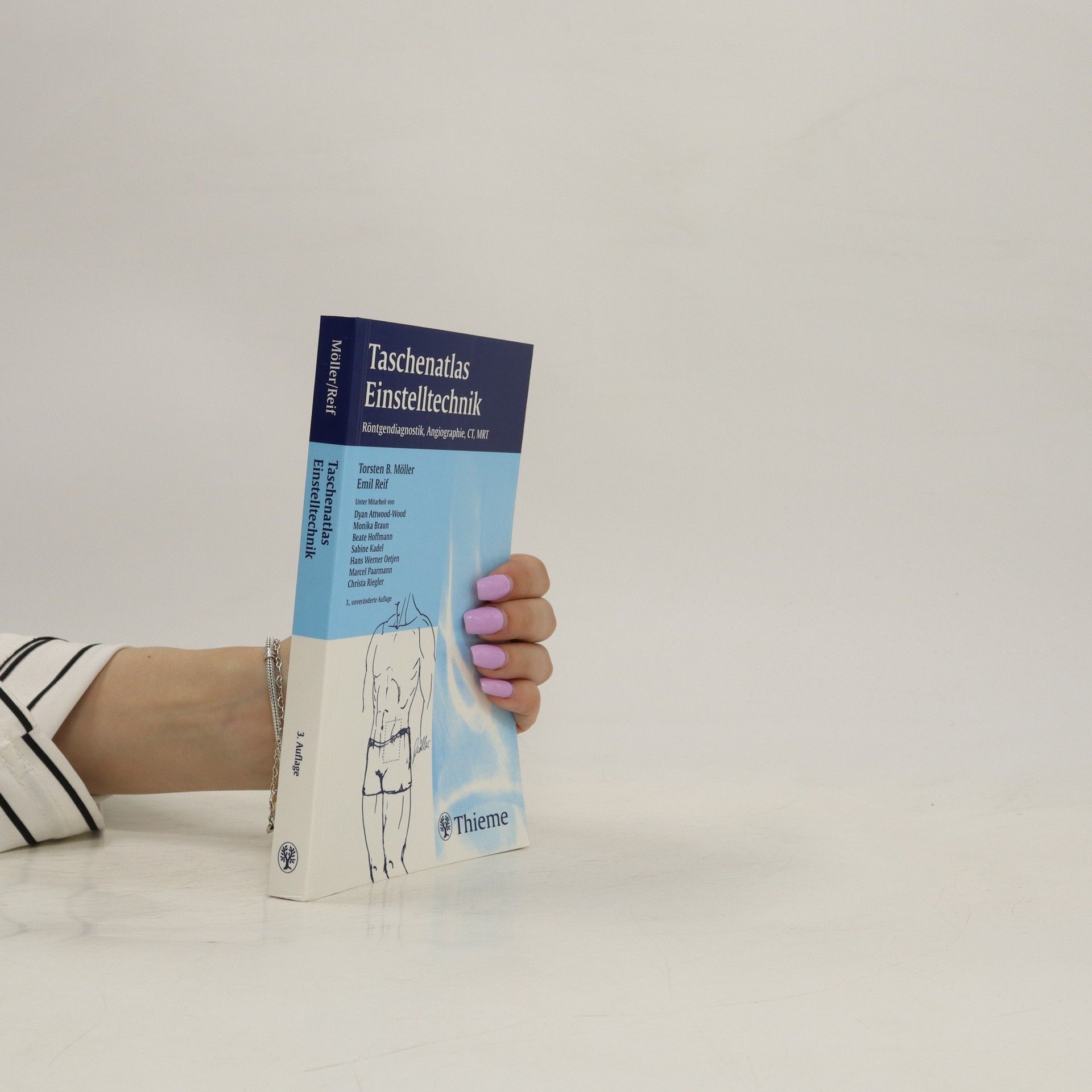
    • Atlas des techniques radiologiques

      • 276pages
      • 10 heures de lecture

      La réalisation correcte d'examens radiologiques est la base d'un diagnostic précis. Une mauvaise incidence ou un film mal exposé peuvent donner de faux résultats. Cet ouvrage s'adresse aux techniciens en radiologie et aux médecins radiologues. Il explique non seulement comment placer le patient, mais également comment obtenir un bon cliché permettant la meilleure interprétation possible. L'ensemble des informations, critères de réussite, " trucs et astuces " sur chaque examen rendent cette tâche plus aisée.

      Atlas des techniques radiologiques
    • L'IRM est devenue un outil essentiel de la pratique quotidienne des radiologistes, orthopédistes et rhumatologistes. Les nouvelles machines plus petites, moins chères, plus faciles à manipuler, vont aider au développement de cette imagerie qui deviendra la méthode diagnostique de choix. Mais on ne peut diagnostiquer un état pathologique sans bien connaître l'aspect du normal. Cet Atlas IRM des membres offre une présentation visuelle exceptionnelle de l'anatomie des muscles et des os. Traitant, en deux sections principales, du membre supérieur et du membre inférieur, il fournit une riche information systématique : chaque page comporte une image IRM accompagnée d'un schéma qui en dérive directement par transfert sur ordinateur. Chaque schéma est légendé et en couleurs codées : les mêmes tissus gardent les mêmes couleurs tout au long de l'ouvrage. Un autre schéma précise l'orientation des coupes. Le caractère systématique de l'information et la clarté de la présentation font de cet Atlas IRM des membres un ouvrage utile, facile à consulter et sans équivalent.

      Anatomie IRM des membres
    • This comprehensive, easy-to-consult pocket atlas is renowned for its superb illustrations and ability to depict sectional anatomy in every plane. Together with its two companion volumes, it provides a highly specialized navigational tool for all clinicians who need to master radiologic anatomy and accurately interpret CT and MR images. Special features of Pocket Atlas of Sectional Anatomy: Didactic organization in two-page units, with high-quality radiographs on one side and brilliant, full-color diagrams on the other Hundreds of high-resolution CT and MR images made with the latest generation of scanners (e. g., 3T MRI, 64-slice CT) Consistent color coding, making it easy to identify similar structures across several slices Concise, easy-to-read labeling of all figuresUpdates for the 4th edition of Volume I: New cranial CT imaging sequences of the axial and coronal temporal bone Expanded MR section, with all new 3T MR images of the temporal lobe and hippocampus, basilar artery, cranial nerves, cavernous sinus, and more New arterial MR angiography sequences of the neck and additional larynx images Compact, easy-to-use, highly visual, and designed for quick recall, this book is ideal for use in both the clinical and study settings.

      Pocket atlas of cross sectional anatomy 1
    • This comprehensive pocket atlas is celebrated for its exceptional illustrations and ability to depict sectional anatomy across all planes. It serves as a specialized navigational tool for clinicians mastering radiologic anatomy and interpreting CT and MR images. Key features include a didactic organization in two-page units, with high-quality radiologic images on one side and vibrant, full-color diagrams on the other. It contains hundreds of high-resolution CT and MR images, many from the latest scanners (e.g., 3T MRI, 64-slice CT), and employs consistent color coding for easy identification of similar structures across slices. The figures are concisely and clearly labeled for ease of understanding. The 4th edition of Volume I includes new cranial CT imaging sequences of the axial and coronal temporal bone and an expanded MR section featuring new 3T MR images of the temporal lobe, hippocampus, basilar artery, cranial nerves, cavernous sinus, and more. It also introduces new arterial MR angiography sequences of the neck and additional images of the larynx. Compact, visually engaging, and designed for quick reference, this atlas is perfect for both clinical and study environments. The volumes cover Head and Neck, Thorax, Heart, Abdomen, Pelvis, Spine, Extremities, and Joints, making it an invaluable resource for medical professionals.

      Pocket atlas of cross sectional anatomy 2
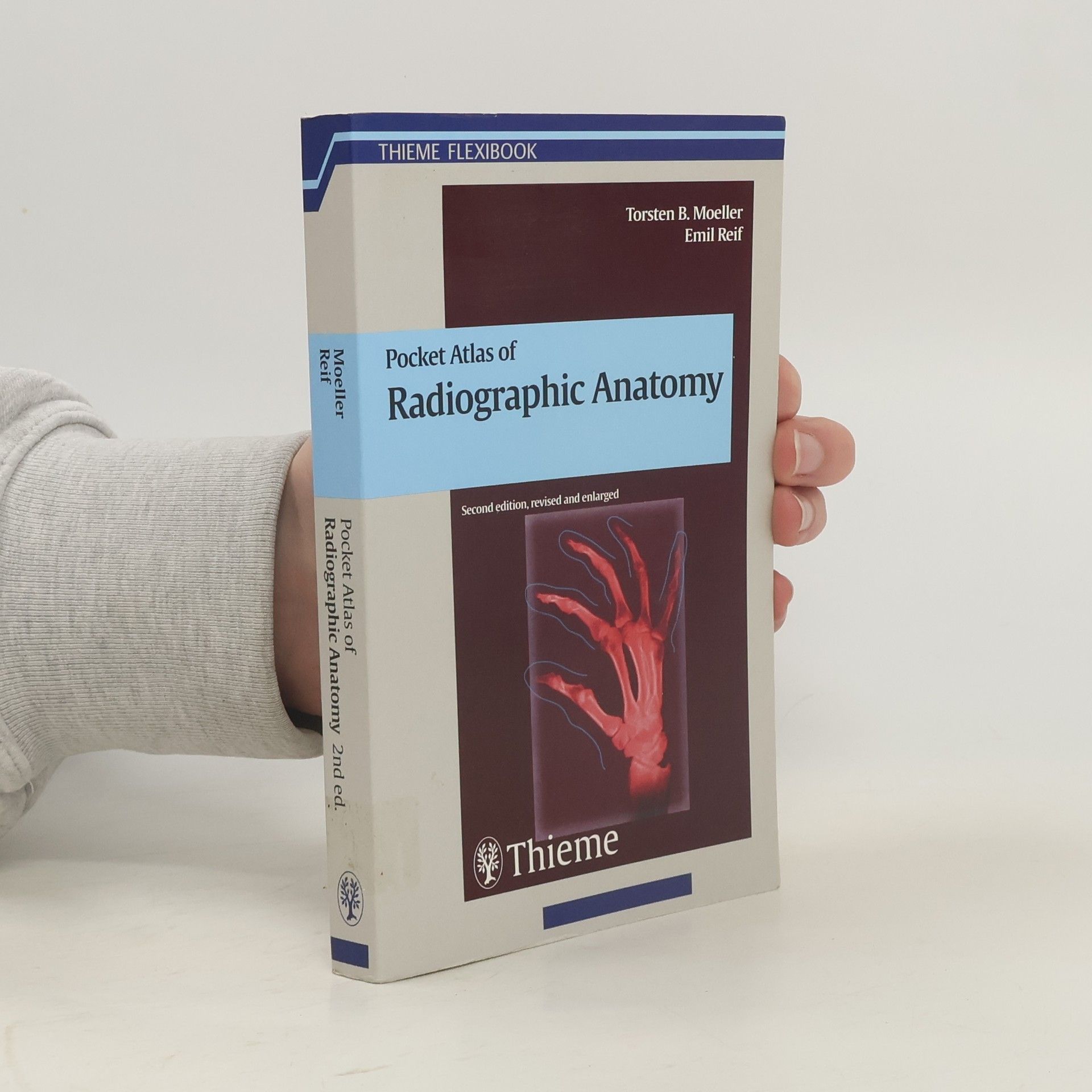
    • Third edition of the classic pocket guide to basic radiographic anatomyIn this easily accessible pocket atlas, two expert radiologists present the normal radiographic anatomy readers need in order to interpret conventional diagnostic radiographs. Each practical, two-page unit displays a standard radiograph of a different projection on the left-hand side supplemented by a detailed, clearly labeled schematic drawing on the opposing page. The consistent, user-friendly format facilitates easy identification and rapid review of key anatomic information. Features:177 radiographic studies provide multiple views of every basic anatomic structure High-resolution radiographs appear beside explanatory drawings to aid comprehension Seven examinations new to this edition cover a trans-scapular „Y“ view of the shoulder; 45° external and internal rotation views of the knee; and moreAn ideal reference for anyone involved in the interpretation of commonly performed radiographic studies, the third edition of Pocket Atlas of Radiographic Anatomy is an especially valuable tool not only for medical students and radiology residents, but also for radiological technologists.

      Pocket atlas of radiographic anatomy
    • MRI parameters and positioning

      • 341pages
      • 12 heures de lecture
      3,3(4)Évaluer

      Packed with information on the practical aspects of MRI, this user-friendly text covers everything from advice on optimal positioning of patients to recommendations for setting the appropriate scanning parameters. Each consistently organized chapter follows the chronology of a standard procedure - the authors present essential information on preparation and necessary materials first. Then they skilfully guide the readers through special considerations in positioning and coil selection, protocols for conducting the exam, examples of various sequences, and possible modifications. Numerous tips, tricks, and pointers explain how to avoid potential complications. Highlights of the second edition: 340 high-quality MRI scans and anatomical drawings New and expanded sections on MR angiography of pulmonary arteries and pelvic and leg vessels; the CARE Bolus Technique; whole-body MRI; and more Information on the latest protocols for MR urography, cholangiography, and colonography Consistent chapter structure for maximum accessibility on the job and at the MRI workstation Each section contains plenty of space on each page for personal notes A guide to the most important MRI studies, the second edition of MRI Parameters and Positioning is an indispensable companion for all radiologists, radiology residents, and radiologic technologists.
      MRI parameters and positioning
    • The second of a two volume set which describes the anatomical details visualized in diagnostic tomography. As a comprehensive reference it is an aid when interpreting images for anatomic structures presented in representative cross-sectional CT and MRI images.

      Pocket Atlas of Cross Sectional Anatomy