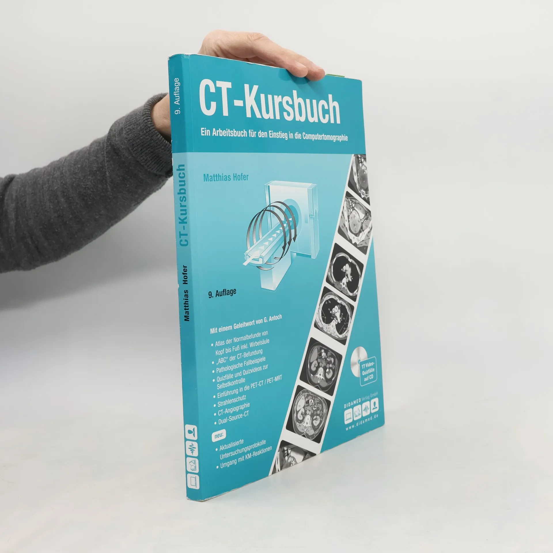
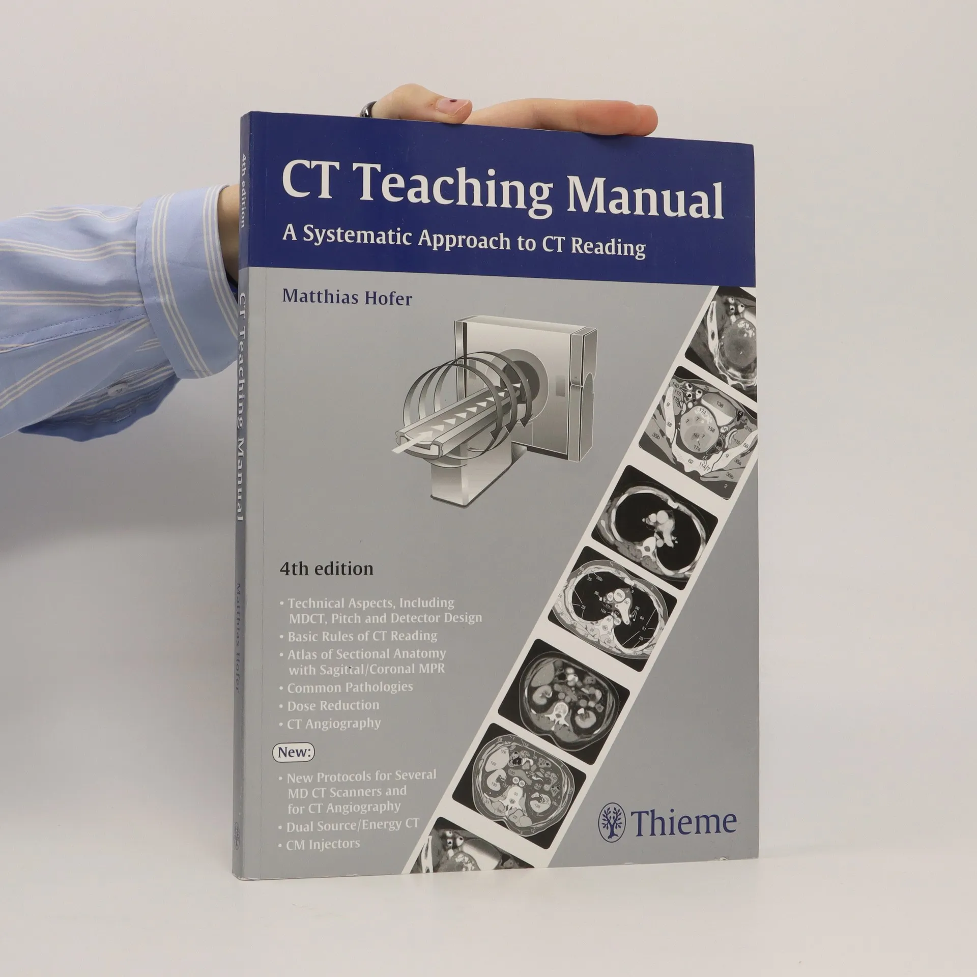
This workbook offers structured, course-like learning, and just like an instructor in an ultrasound course, it guides you systematically through the individual organ systems. The accompanying videos demonstrate basic anatomy for ultrasound, optimum transducer positioning, and the interaction between transducer position and monitor display, allowing you to experience the learning points in real time for a deeper, visual understanding
For whom is this book designed? For all students and physicians in training
who want to learn more about the systematic interpretation of conventional
chest radiographs, and for anyone who wants to learn how to insert chest tubes
and central venous catheters. What does this book offer? - Detailed diagrams
on topographical anatomy, with numerical labels for self-review.- Coverage
includes even relatively complex findings in trauma victims and ICU patients.-
Detailed, step-by-step instructions on the placement of CVCs and chest tubes.-
Simple aids and tricks, such as the silhouette sign, that are helpful in image
interpretation.- Images to illustrate all common abnormalities (systematically
arranged according to morphological patterns).
Ideal for residents starting in radiology and radiologic technologists, this concise manual is the perfect introduction to the physics and practice of CT and the interpretation of basic CT images. Designed as a systematic learning tool, it introduces the use of CT scanners for all organs, and includes positioning, use of contrast media, representative CT scans of normal and pathological findings, explanatory drawings with keyed anatomic structures, and an overview of the most important measurement data. Finally, self-assessment quizzes - including answers - at the end of each chapter help the reader monitor progress and evaluate knowledge gained. New in this fourth edition: - Updated examination protocols for multidetector CT and CT angiography - An extended chapter on dual source CT - A new chapter on contrast injectors
The Third Edition of this up-to-date and user friendly workbook helps medical students, sonographers, residents, and radiologists gain a fundamental grasp of the application of color duplex ultrasound by reviewing normal findings, important pathologic conditions, scanning techniques, and the role and importance of color duplex ultrasound in detecting and assessing various disease states. Topics include: Basic physical and technical principles Innovative techniques and ultrasound contrast agents (e. g., power Doppler, SieScape imaging, clarify vascular enhancement, tissue Doppler, precision upsampling, arterial stiffness, eTracking) Vascular surgery: peripheral arterial occlusive disease, venous insufficiency and thrombosis, AV fistulae, and aneurysms Endocrinology: thyroid gland Internal medicine: abdominal organs, lymph nodes, TIPSS Nephrology: kidneys and renal allografts Neurology: intra- and extracranial cerebral arteries Cardiology: B- and M-mode imaging, cardiac anomalies, wall motion analysis Urology: testicular torsion, tumors, erectile dysfunction Obstetrics and gynecology: tumors, anomalies, fetal perfusion defectsTeaching Manual of Color Duplex Sonography is well known for its highly educational approach and user-friendly features which include: Over 500 excellent illustrations -- ideal for visually oriented learners Diagrams that illustrate the correct way to handle the transducer and position the scan plane Self-test question & answers Tips, tricks, and common pitfalls Checklists and tables of normal values on sturdy pocket-sized cards for hospital and office use
Explore the evolution of the German language in Austria with this groundbreaking study by Matthias Höfer. Focusing on the unique features of the Austrian dialects, Höfer offers insights into the ways in which language reflects the cultural and historical context in which it develops. A must-read for linguists, historians, and anyone interested in the fascinating history of this complex and diverse region.
Studienarbeit aus dem Jahr 2004 im Fachbereich Germanistik - Linguistik, Note: 5-6 (CH), Universität Zürich (Deutsches Seminar der Universität Zürich), Veranstaltung: Proseminar: Einführung in die Pragmatik, Sprache: Deutsch, Abstract: Abstract Der Text behandelt eine metalinguistische Debatte, die [im Grunde bereits /MH] seit Beginn des 19. Jahrhunderts die Geschichte der Linguistik durchzogen hat (Jäger 1993a: 77), die Jäger-Bierwisch-Grewendorf-Debatte . Der Disput wird zwischen Funktionalisten und Strukturalisten (Mead vs. Chomsky) geführt. Er geht der Frage nach dem Untersuchungsgegenstand der Linguistik nach. In einem ersten Teil soll die Debatte zwischen Ludwig Jäger er tritt auf als vehementer Verteidiger von funktionsorientierten Theorien auf der einen sowie Manfred Bierwisch und Günther Grewendorf zwei generativen Grammatikern auf der anderen Seite vorgestellt werden. In einem zweiten Teil wird zu beiden Positionen kritisch Stellung genommen, wobei mittels prak¬tischer Beispiele versucht wird, die Leistungsfähigkeit und die Art und Weise der beiden Herange¬hensweisen an das Phänomen Sprache deutlich zu machen. Am Ende wird versucht eine Synthese zu formulieren.
Das Buch ist nicht nach Krankheitsgruppen, sondern nach bildmorphologischen „Symptomen“, wie z. B. einer Mediastinalverbreiterung oder flächiger Lungenveränderungen gegliedert. Innerhalb eines Kapitels sind dann die pathologischen Ursachen nach Häufigkeit mit Fallbeispielen und detaillierten anatomischen Skizzen erläutert. Spannende Quizfälle zum Selbsttest im Buch und zusätzliche Videoclips mit Quizbeispielen sind online abrufbar. Neu ist ein Kapitel zu Corona-induzierten Lungenveränderungen im zeitlichen Verlauf.