

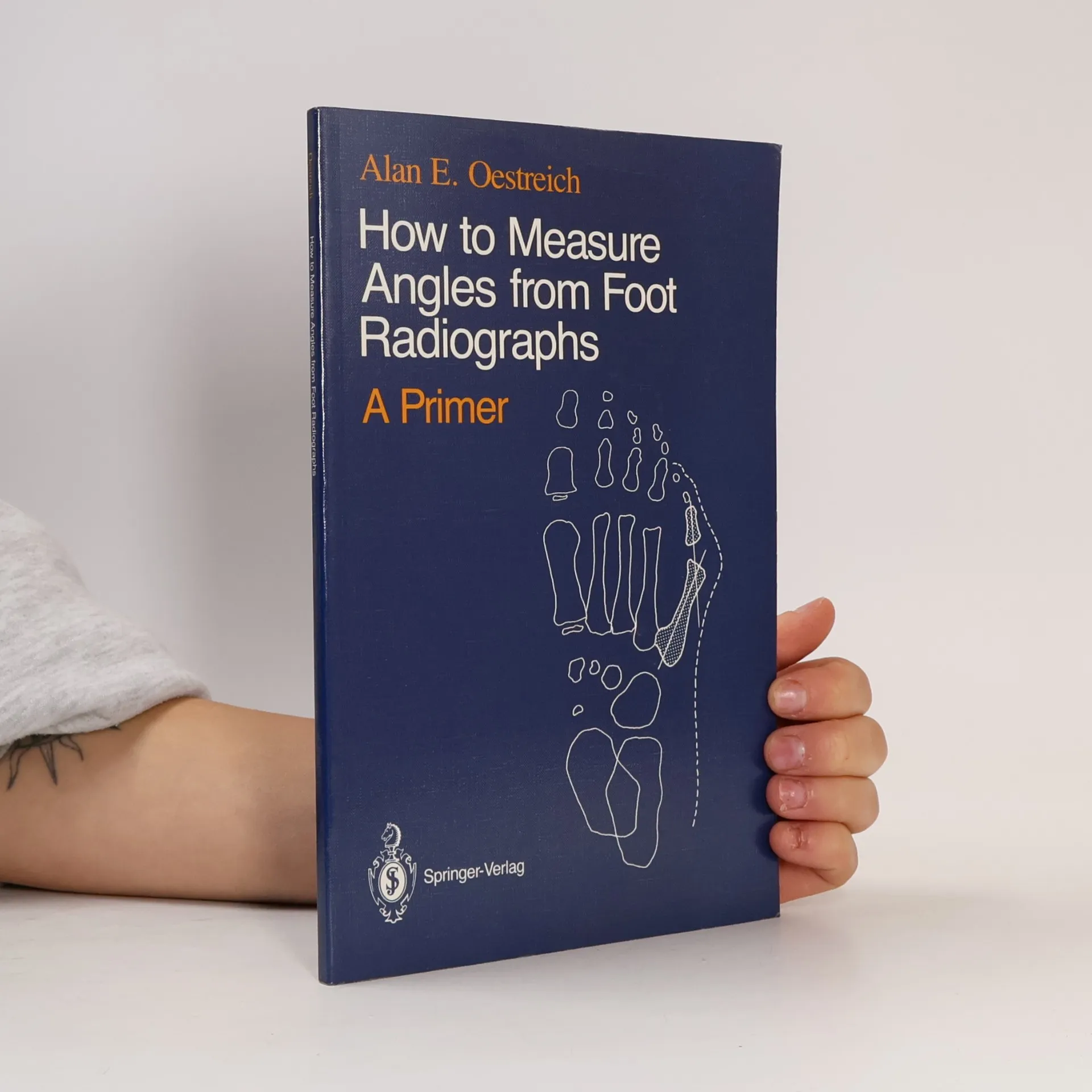
How to Measure Angles from Foot Radiographs
A Primer
En savoir plus sur le livre
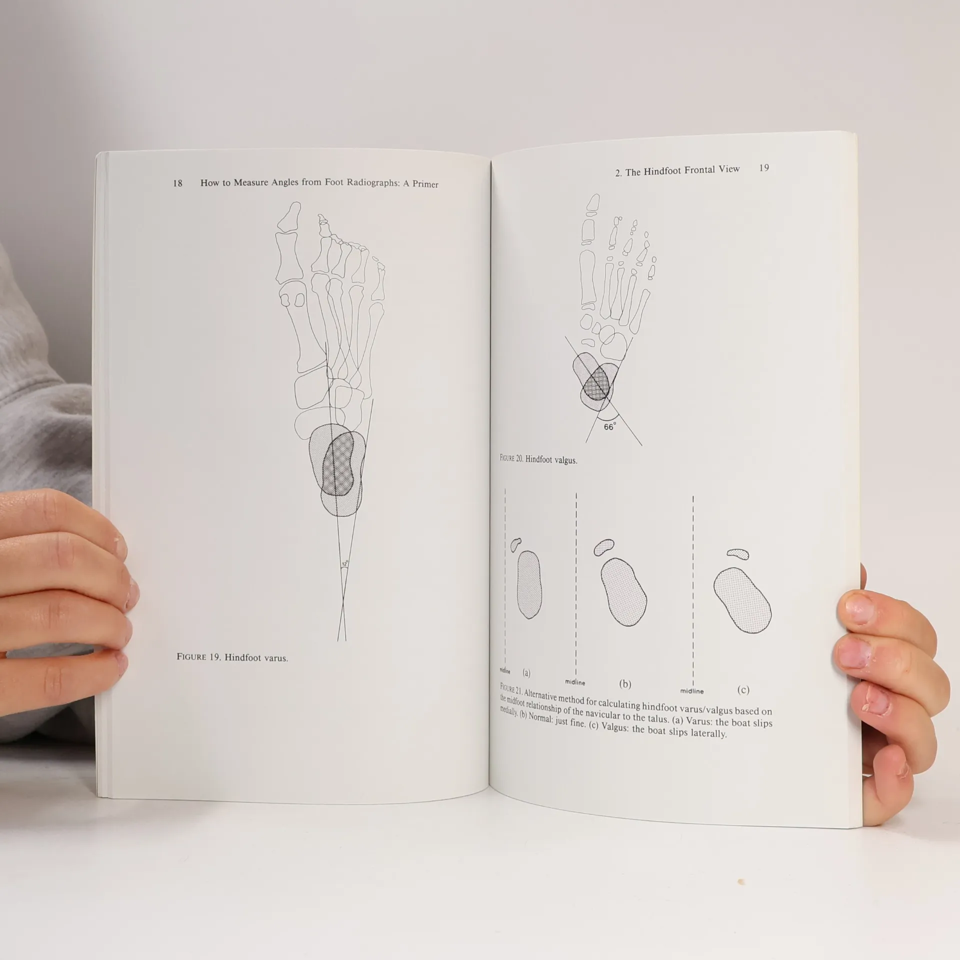
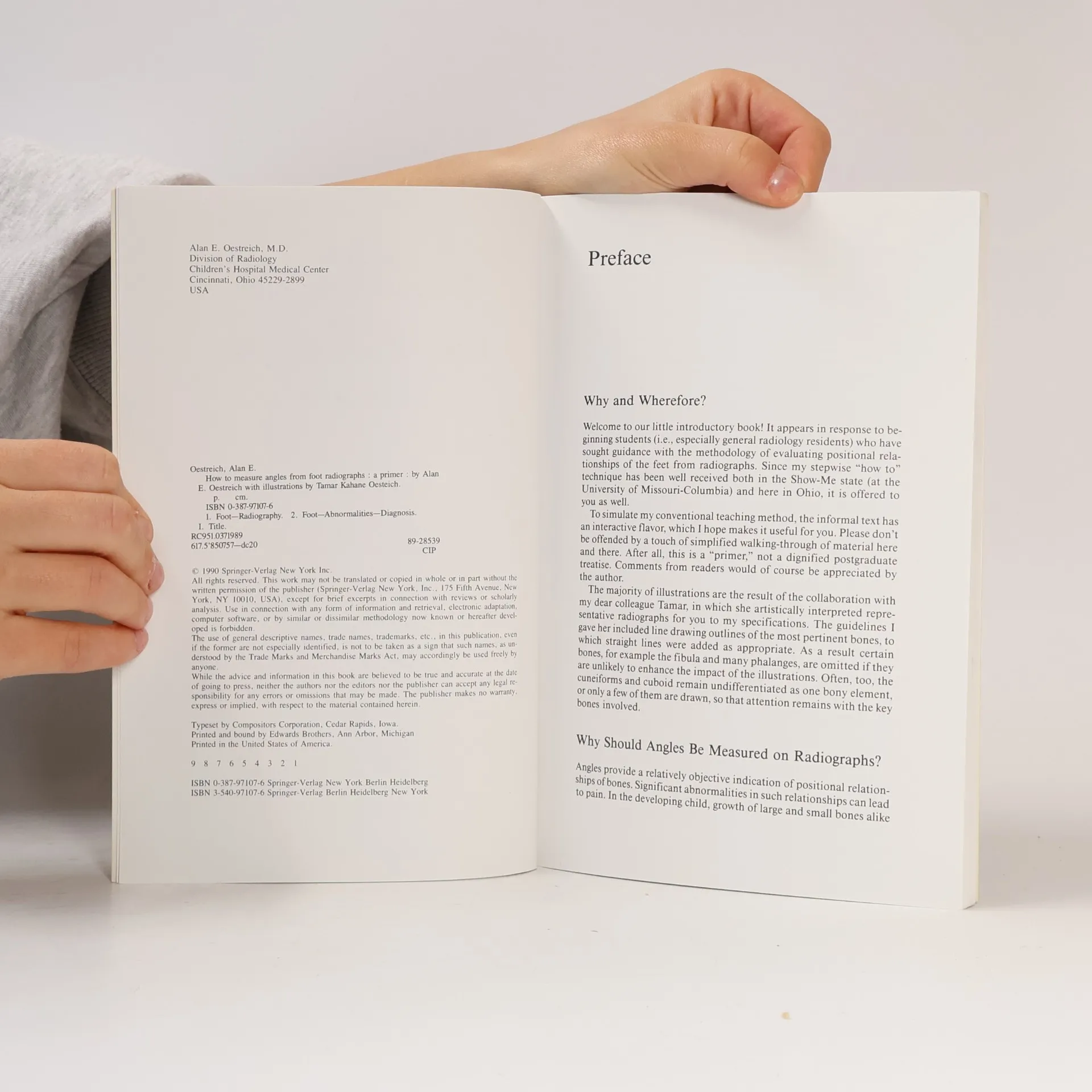
Why and Wherefore? Welcome to our little introductory book! It appears in response to be ginning students (i.e., especially general radiology residents) who have sought guidance with the methodology of evaluating positional rela tionships of the feet from radiographs. Since my stepwise "how to" technique has been well received both in the Show-Me state (at the University of Missouri·Columbia) and here in Ohio, it is offered to you as well. To simulate my conventional teaching method, the informal text has an interactive flavor, which I hope makes it useful for you. Please don't be offended by a touch of simplified walking·through of material here and there. After all, this is a "primer," not a dignified postgraduate treatise. Comments from readers would of course be appreciated by the author. The majority of illustrations are the result of the collaboration with my dear colleague Tamar, in which she artistically interpreted repre sentative radiographs for you to my specifications. The guidelines I gave her included line drawing outlines of the most pertinent bones, to which straight lines were added as appropriate. As a result certain bones, for example the fibula and many phalanges, are omitted if they are unlikely to enhance the impact of the illustrations. Often, too, the cuneiforms and cuboid remain undifferentiated as one bony element, or only a few of them are drawn, so that attention remains with the key bones involved.
Achat du livre
How to Measure Angles from Foot Radiographs, Tamar K. Oestreich, Alan E. Oestreich
- Langue

- Année de publication
- 1990
- Reliure
- (souple),
- État du livre
- Bon
- Prix
- 40,99 €
Modes de paiement
Personne n'a encore évalué .
- Titre
- How to Measure Angles from Foot Radiographs
- Sous-titre
- A Primer
- Langue
- Anglais
- Auteurs
- Tamar K. Oestreich, Alan E. Oestreich
- Éditeur
- Springer
- Publié
- 1990
- Format
- souple
- Pages
- 53
- ISBN10
- 0387971076
- ISBN13
- 9780387971070
- Séries
- Catégories
- Mots clés
- Médecine, Littérature spécialisée, Chirurgie, Biochimie, Méthodologie, Radiologie, Orthopédie, Médecine nucléaire, Podologie
- Description
- Why and Wherefore? Welcome to our little introductory book! It appears in response to be ginning students (i.e., especially general radiology residents) who have sought guidance with the methodology of evaluating positional rela tionships of the feet from radiographs. Since my stepwise "how to" technique has been well received both in the Show-Me state (at the University of Missouri·Columbia) and here in Ohio, it is offered to you as well. To simulate my conventional teaching method, the informal text has an interactive flavor, which I hope makes it useful for you. Please don't be offended by a touch of simplified walking·through of material here and there. After all, this is a "primer," not a dignified postgraduate treatise. Comments from readers would of course be appreciated by the author. The majority of illustrations are the result of the collaboration with my dear colleague Tamar, in which she artistically interpreted repre sentative radiographs for you to my specifications. The guidelines I gave her included line drawing outlines of the most pertinent bones, to which straight lines were added as appropriate. As a result certain bones, for example the fibula and many phalanges, are omitted if they are unlikely to enhance the impact of the illustrations. Often, too, the cuneiforms and cuboid remain undifferentiated as one bony element, or only a few of them are drawn, so that attention remains with the key bones involved.